Month: April 2018

New Clinical Data for Covalon’s ColActive®

Line of Patented Advanced Wound Care Products to be Presented at SAWC Spring


MISSISSAUGA, Ontario–(BUSINESS WIRE)–Covalon Technologies Ltd. (the “Company” or “Covalon”) (TSXV: COV), an advanced medical technologies company, today announced that two clinical case studies demonstrating the clinical effectiveness of ColActive® Plus and ColActive® Transfer biological matrix products will be presented at the 2018 Symposium on Advanced Wound Care Spring (“SAWC Spring”) in Charlotte, North Carolina. In addition to the posters, Covalon will also be showcasing its full line of advanced wound care dressings in the symposium’s exhibition hall during the three-day event.


“We are very pleased with the results of these two clinical case studies and believe that they show the advantage that the patented ColActive technology can add to a wound care clinician’s arsenal,” said Brian Pedlar, Chief Executive Office of Covalon.


Dr. Jeffrey Lehrman, DPM, FASPS, MAPWCA will present a clinical poster entitled A New Approach in the Management of Chronic Diabetic Foot Ulcers: A Report on the Use of a Collagen Wound Contact Layer with Negative Pressure Wound Therapy during the SAWC Spring – WHS Poster Gala, held on Friday, April 27th from 7:15 p.m. to 8:45 p.m.

read more

Wound Care Advantage Launches Advanced Discovery Research Alliance

SIERRA MADRE, Calif.–(BUSINESS WIRE)–Apr 24, 2018–Wound Care Advantage (WCA), in collaboration with internationally renowned podiatric surgeon Dr. David G. Armstrong, the Southwestern Academic Limb Salvage Alliance (SALSA) and Gen1 Research, has launched Advanced Discovery, a new investigative research alliance dedicated to revolutionizing patient care through evidence-based science. Advanced Discovery will offer access to an international network of advanced wound and hyperbaric treatment centers, laying the groundwork for meaningful investigative research. In partnership with approved manufacturers and research facilitators, the Alliance will evaluate a variety of diagnostic and treatment modalities with a unified goal of positively impacting wound healing outcomes while reducing the cost of treatment. Gen1 will provide extensive administrative support and infrastructure for the studies.


Chronic wounds affect approximately 5.7 million patients in the U.S., costing the healthcare system more than $20 billion annually. 1 This burden is growing, due to a rapid increase in diabetes and vascular disease, an aging population, and rising healthcare costs. In fact, the cost of diabetic foot ulcers is greater than that of the five most costly forms of cancer 2 and diabetic foot ulcer patients are twice as costly to U.S. Medicare as those with diabetes alone … read more

Organogenesis Showcases New Product Offerings

and Research at SAWC Spring 2018


CANTON, Mass. and CHARLOTTE, N.C., April 24, 2018 /PRNewswire/ — The latest advanced wound care research and product offerings from Organogenesis Inc. – including the recently-launched PuraPly® Antimicrobial 1.6 cm small size – will be showcased during the nation’s largest interdisciplinary wound care forum, the Symposium on Advanced Wound Care Spring | Wound Healing Society (SAWC Spring | WHS) meeting, held April 25-29 in Charlotte, NC.


Organogenesis Inc., a leading regenerative medicine company focused on the development, manufacture and commercialization of product solutions for the advanced wound care, surgical and sports medicine markets, will feature its full suite of advanced wound care and surgical biologic product offerings at exhibit booth #419.


Scientific presentations will feature several products within the Organogenesis portfolio, and exhibit booth attendees will have the chance to learn more about the company’s comprehensive wound care portfolio designed to empower personalized healing for a wide variety of wound types across the wound care continuum.


Organogenesis is also a proud sponsor of the Thursday, April 26 Lunch Symposium, “Understanding the Latest Evidence: A Fresh Look at the Use of Skin Substitutes Across the Wound Care Continuum,” featuring speakers Daniel Kapp, MD; George Koullias, MD; and Katie C. Mowry, PhD.

Original Press Release on PR Newswire

Effectiveness of using a new polyurethane foam multi-layerdressing

in the sacral area to prevent the onset of pressure ulcerin the elderly with hip fractures: A pragmatic randomisedcontrolled trial


Hip fractures in the elderly are a serious problem for the health service due to the highrate of complications. One of these complications is pressure ulcers that, according to the literature, occur in 8.8% to 55% of patients and mainly arise in the sacral area.The present randomised controlled trial tests whether applying a new innovativemulti-layer polyurethane foam dressing (ALLEVYN LIFE™), reduces the onset ofpressure ulcers in the sacral area. From March to December 2016, 359 fragility hipfracture patients were randomly divided into 2 groups: 182 in the control group and177 in the experimental group. Pressure ulcers occurred overall in 36 patients (10%):8 patients (4.5%) in the experimental group compared to 28 (15.4%) in the controlgroup:P= 0.001, relative risk 0.29 (95% CI 0.14-0.61) with NNT of 9 (95% CI 6-21). In the experimental group the onset of pressure ulcers occurred on average onthe 6th day compared to the 4th day in the control group (HR 4.4). Using polyure-thane foam is effective at reducing the rate of pressure ulcers in the sacrum in elderlypatients with hip fracture. The adhesiveness of this device also enables costs to be kept down … read more

Protecting Periwound Skin in Chronic Wounds

The periwound is as important as the wound. As clinicians, we should carefully assess the wound bed, but we need to remember also to assess the periwound and surrounding skin. The periwound should be considered the 4cm of surrounding skin extending from the wound bed. Chronic wounds may manifest any of the following characteristics, depending on wound type: erythema, induration, epibole, ecchymosis, hyperkeratosis, and changes in shape.

Five-Step Periwound Assessment

  1. Temperature
  2. Location
  3. Shape
  4. Color
  5. Wound depth

The temperature of the periwound can be a good indicator of whether active infection is present or to determine whether there is normal blood flow. The back of the hand is most accurate … read more

Medline to Highlight Holistic Approach to Wound Care

and Skin Health at Symposium on Advanced Wound Care


NORTHFIELD, Ill.–(BUSINESS WIRE)–Medline today announced the launch of a new Skin Champion Program at this year’s Symposium on Advanced Wound Care (SAWC) in Charlotte, April 25-29. With more than 6.5 million people in the U.S. suffering from chronic wounds, the Medline Skin Champion Program will provide new approaches to those who battle and treat non-healing wounds across the continuum of care and provide easy-to-use education and tools for wound care clinicians and leaders.


Medline also will host a live studio news show from SAWC, featuring nearly 20 key advanced wound care and tissue regeneration opinion leaders who will provide clinical expertise and insight to the more than 2,000 physicians and nurses visiting the conference.


“Wound care has become increasingly complex with the rise in patient acuity and ongoing reimbursement challenges,” says Jonathan Primer, group president, advanced wound care, Medline. “For clinicians, this new landscape is creating greater demand for education and fueling a deeper desire to discover new ways to improve clinical practice and patient care.”


Located at booth 1025, Medline will unveil the new Skin Champion Program developed by certified WOC nurses and designed to provide education and tools that help train their teams to make skin health second nature. Dozens of pre-built educational modules address four common issues impacting wound care, including pressure injuries, skin care, wound etiologies and special populations.

read more

Healogics, Inc. Names Allan Woodward

MBA as New Chief Financial Officer


JACKSONVILLE, Fla.–(BUSINESS WIRE)–Healogics, the nation’s largest provider of advanced chronic wound care services, today announced that it has appointed Allan Woodward to serve as Chief Financial Officer. In this role, Woodward will be responsible for the planning and oversight of the financial strategy for the company, the ongoing development of appropriate control systems and all reporting measures. Woodward will report directly to the Chief Executive Officer, David Bassin.


“Allan’s extensive experience in financial leadership roles, combined with his knowledge of the healthcare industry makes him an invaluable asset to our team. We are thrilled to welcome him as our new CFO,” said Bassin.


Over the course of his career, Allan has held several financial leadership roles in healthcare-based organizations. His most recent experience includes his service at eviCore Healthcare as the Senior Vice President of Finance and Strategy over the post-acute care and consumer engagement programs. He was responsible for all financial activities of the business unit, including the creation of financial structures for all product expansions and the development of alternative provider reimbursement initiatives.


Woodward earned his MBA from the University of Missouri and his Bachelor of Science from Middle Tennessee State University … read more

MiMedx to Highlight Efficacy of Its Allografts

in Presentations at SAWC Spring Conference
Breakfast Symposium to Underscore Statistically Significant Evidence of EpiFix® in Healing DFUs and VLUs


MARIETTA, Ga.April 23, 2018 /PRNewswire/ — MiMedx Group, Inc. (NASDAQ: MDXG), a leading developer and marketer of regenerative and therapeutic biologics, today announced the Company’s participation in the 2018 Symposium on Advanced Wound Care Spring | Wound Healing Society (SAWC Spring/WHS) meeting being held April 25-29, 2018 in Charlotte, North Carolina.

During the five-day conference, the clinical and cost-effective healing results of the Company’s allografts will be highlighted in a Breakfast Symposium, two dinner presentations, five clinical poster presentations, and in-booth education sessions. MiMedx will be exhibiting in booth #830 from April 26, 2018 through April 28, 2018.


Now in its 31st year, the SAWC Spring/WHS conference is the leading national wound healing forum connecting the foremost experts with your entire wound care team to improve patient outcomes through education.  No other wound care conference offers the level of education, advanced state-of-the-art clinical reviews and emerging research findings.


MiMedx will sponsor a Breakfast Symposium entitled “EpiFix® – First and Only Amnion/Chorion Allograft with Statistically Significant Level I Evidence for healing in DFUs and VLUs” on Friday morning, April 27, 2018 from 7:30 am to 9:00 am. The Company will also host two dinner presentations on Thursday, April 26th and Friday, April 27th entitled “Using MiMedx Placental Tissues in the Lower Extremity“, which will review the use of various products in wound and surgical applications.


Hands-on product demonstrations will be provided by expert physicians during booth hours on April 26th and April 27thin the MiMedx booth.


The poster presentations will report on the clinical effectiveness of MiMedx EpiFix dHACM (dehydrated Human Amnion/Chorion Membrane) placental tissue allografts in the healing of diabetic foot ulcers, venous leg ulcers, pressure ulcers and refractory non-healing wounds, as well as the use of AmnioCord umbilical cord allografts for reducing Achilles tendon pain. These independent case studies and respective conclusions will include:


  • Abstract: “EpiFix VLU Study Group: A Multicenter Randomized Controlled Trial Evaluating the Efficacy of Dehydrated Human Amnion/Chorion Membrane Allograft for the Treatment of Venous Leg Ulcers
    Authors: Christian Bianchi, MD, FACS; Shawn Cazzell, DPM, FACFAS; Dean Vayser, DPM, FACFAS; Alexander M. Reyzelman, DPM, FACFAS; Hasan Dosluoglu, MD, FACS; Gregory Tovmassian, DPM
  • Abstract: “EpiFix DFU Study Group: A Confirmatory Study on the Efficacy of Dehydrated Human Amnion/Chorion Membrane (dHACM) Allograft in the Management of Diabetic Foot Ulcers: A Prospective, Multicenter, Randomized, Controlled Study
    Authors: William Tettelbach, MD; Shawn Cazzell, DPM; Alexander M. Reyzelman, DPM; Felix Sigal, DPM; Joseph M. Caporusso, DPM; Patrick S. Agnew, DPM
  • Abstract: “The Application of Dehydrated Human Amnion Chorion Membrane dHACM Allografts to Expedite Healing in Patients with Six Major Types of Refractory Non-Healing Wounds, 157 Patients
    Authors: Aamir Mahmood, DPM; Justin Goldsmith, DPM; Anna Tien, DPM; Mike Czurylo, DPM; Laith Shaman, DPM; Kelda Beachy, DPM; Neal Patel, DPM; Shayan Alamgir, DPM; Matthew Garoufalis, DPM, FASPS, FACFAOM
  • Abstract: “Use of Umbilical Cord Allograft for Pain Reduction in Achilles Tendon Pathologies: A Case
    Authors: Brandon Brooks, DPM; Kevin Pham, DPM; Bradley Brooks, DO; Brady Brooks, MS-1; James Henry, MS-1; Terria Madison, DPM
  • Abstract: “Safety and Efficacy of Weekly Application of Dehydrated Human Amnion/Chorion Membrane in the Treatment of Pressure Ulcers: a Case Series
    Authors: Chi Chi Berhane, MD, MBA; Kimberly Brantley, BSN, RN, CRRN; Sandra Williams, NP-C, APN, WCN; Erica Sutton, MACarlyn Kappy, RD, LD, CCRP


“For more than 30 years, SAWC and WHS have been dedicated to continuous advancements in wound care and have worked tirelessly toward their goal of decreasing the number and severity of wounds of all types.” said Parker H. “Pete” Petit, MiMedx Chairman and CEO. “We are honored to be a part of this year’s meeting, and MiMedx will continue to work with SAWC and other organizations to raise the level of scientific and clinical expertise and the professional processes within the wound care sector of healthcare.”


Bill Taylor, President and COO of MiMedx, added, “We are pleased that our EpiFix, AmnioCord and AmnioFill allografts will be so prominently demonstrated at this year’s SAWC. We look forward to participating in the conference and highlighting the significant investments made in the science and clinical study of placental technology.”


About MiMedx


MiMedx® is a leading biopharmaceutical company developing and marketing regenerative and therapeutic biologics utilizing human placental tissue allografts with patent-protected processes for multiple sectors of healthcare. “Innovations in Regenerative Medicine” is the framework behind the Company’s mission to give physicians products and tissues to help the body heal itself.  The Company processes the human placental tissue utilizing its proprietary PURION® Process methodology, among other processes, to produce safe and effective allografts by employing aseptic processing techniques in addition to terminal sterilization. MiMedx has supplied over 1 million allografts to date for application in the Wound Care, Burn, Surgical, Orthopedic, Spine, Sports Medicine, Ophthalmic and Dental sectors of healthcare. For additional information, please visit www.mimedx.com.

Press release from PR Newswire

An evaluation of an ultrasonic debridement system

in patients with diabetic foot ulcers: a case series


Ferdinando CampitielloMD , Manfredi ManconeMD , Angela Della CorteMD , Raffaella GuernieroMD , Silvestro Canonico


Objective:
This study evaluated the use of ultrasonic debridement in patients with diabetic foot ulcers (DFU).


Method:
In this prospective, single-arm, open-label study, all patients with DFUs underwent wound debridement by ultrasonic debridement system (SonicOne OR Ultrasonic debridement system). Wherever possible, the edges were approximated by means of stitches. In other cases, the surgical breach healed by secondary intention, or a partial thickness skin graft (with or without Integra Dermal Regeneration Template or Integra Flowable Wound Matrix) was applied, and subsequently healed by primary intention … read more

Sucrose octasulfate wound dressing may speed healing in diabetic foot ulcers

Patients with a noninfected neuroischemic diabetic foot ulcer were more likely to achieve wound closure within 20 weeks when randomly assigned a sucrose octasulfate dressing vs. those assigned a standard wound dressing, according to findings published in The Lancet Diabetes & Endocrinology.


“Delayed wound healing in neuroischemic diabetic foot ulcers has been related to excess matrix metalloprotease concentrations; these proteins destroy components of the extracellular matrix and damage growth factors and their receptors that are essential for healing,” Michael Edmonds, MD, of the Diabetic Foot Clinic at King’s College Hospital in London, and colleagues wrote. “Sulfated oligosaccharides are known to have many biological activities; in particular, the potassium salt of sucrose octasulfate has been shown to inhibit matrix metalloproteases and to interact with growth factors and restore their biological functions because it has high charge density.”


Edmonds and colleagues analyzed data from 240 patients with diabetes and a noninfected neuroischemic diabetic foot ulcer of grade IC or IIC, with a wound  … read more

To evaluate the efficacy of an acellular Flowable matrix

in comparison with a wet dressing for the treatment of patients with diabetic foot ulcers: a randomized clinical trial


The authors aimed to evaluate the efficacy of an advanced wound matrix (Integra Flowable Wound Matrix, Integra LifeScience Corp, Plainsboro, NJ, USA) for treating wounds with irregular geometries versus a wet dressing in patients with diabetic foot ulcers. Sixty patients with diabetic foot ulcers (Grades 3 Wagner) were included in this randomized clinical trial. The study was conducted in the General Surgery Unit and Geriatric of the Second University of Naples, Italy, in the last 12 months. Forty-six cases of diabetic foot ulcers were equally and randomly divided into control and test groups. The first group treated with Integra Flowable Wound Matrix, while the control group with a wet dressing. Both groups were evaluated once a week for 6 weeks to value the degree of epithelialization and granulation tissue of the wound. The complete healing rate in the whole study population was 69.56% (Integra Flowable Wound Matrix group, 86.95%, control group, 52.17%; p = 0.001). Amputation and rehospitalization … read more

New hope for treating diabetic wounds that just won’t heal

Mice bred without TSP2 protein heal faster, suggesting a new target for better treatments


One of the most frustrating and debilitating complications of diabetes is the development of wounds on the foot or lower leg. Once they form, they can persist for months, leading to painful and dangerous infections.


New research uncovers the role of a particular protein in maintaining these wounds and suggests that reversing its effects could help aid wound healing in patients with diabetes.


“We discovered that a specific protein, thrombospondin-2 (TSP2), is elevated in wounds of patients with diabetes as well as in animal models of diabetes,” said Britta Kunkemoeller, a doctoral student at Yale University who conducted the study. “To determine whether TSP2 contributes to delayed wound healing, we genetically removed TSP2 from a mouse model of diabetes and observed improved wound healing. Our study shows that TSP2 could be a target for a specific therapy for diabetic wounds.” … read more

Lower Extremity Amputation and Reamputation Predictors

Temple University School of Podiatric Medicine Journal Review Club
Editor’s note: This post is part of the Temple University School of Podiatric Medicine (TUSPM) journal review club blog series. In each blog post, a TUSPM student will review a journal article relevant to wound management and related topics and provide their evaluation of the clinical research therein.

Article Title: Predictors of Lower Extremity Amputation and Reamputation Associated With the Diabetic Foot
Authors: Erdinc Acar MD, Burkay Kutluhan Kacıram, MS
Journal name and issueThe Journal of Foot & Ankle Surgery 56 (2017) 1218–1222
Reviewed by: Anthony Samaan, Class of 2018, Temple University School of Podiatric Medicine

Introduction

A major concern in managing patients with diabetes is their susceptibility to acquiring ulcers in their feet. If these patients are not careful, these ulcers may become infected and eventually lead to additional sequelae, ending in lower extremity amputation. The focus of this study was to determine the major factors of lower extremity amputation in the diabetic foot, in hopes that clinicians may be able to reduce the rate of amputations more effectively.

Methods

The authors performed a retrospective review of the records of 132 consecutive patients who had already received a lower extremity amputation or reamputation as a result of diabetic wounds. All patients had been diagnosed with diabetes mellitus type 2, and demographic and clinical data were collected on all of them. These data included age, sex, cigarette smoking history, duration of diabetes, diabetic comorbidities (nephropathy, neuropathy), general comorbidities (peripheral artery disease, hypertension, hyperlipidemia, malignancy), leukocytosis, wound infection status, and culture microorganism and antibiogram results. The side and level of amputation or reamputation were also recorded. Only those patients with wounds of a Wagner-Meggitt classification of 3 to 6 were included …  read more

HbA1c, wound healing unrelated in diabetic foot ulcers

Among patients with long-term diabetic foot ulcers, neither baseline HbA1c nor change in HbA1c was associated with wound healing time, according to findings from a clinic-based observational study.


“Although we know that chronic hyperglycemia leads to neuropathy and peripheral vascular disease, which are the proximal risk factors for diabetic foot ulcers, we did not see a clear association between HbA1c levels and wound healing in patients who have developed foot ulcers,” Nestoras Mathioudakis, MD, MHS, assistant professor of medicine and clinical director, division of endocrinology, diabetes and metabolism at Johns Hopkins University School of Medicine, told Endocrine Today. “It is likely that the damage induced by chronic hyperglycemia reaches a point where it cannot be reversed in a relatively short time frame to improve wound healing.”

read more

B. Braun to Introduce 1-Ounce Prontosan® Wound Gel X at SAWC

BETHLEHEM, Pa., April 19, 2018 (GLOBE NEWSWIRE) — B. Braun Medical Inc. will introduce a patient-friendly 1-ounce tube of Prontosan®Wound Gel X at the 2018 Symposium of Advanced Wound Care, which is being held April 26-28 in Charlotte, North Carolina.


A viscous wound gel with both betaine and polyhexanide to resist microbial colonization within the dressing, Gel X softens necrotic tissue, facilitating autolytic debridement. Clear and virtually odorless, Gel X is designed to cleanse and moisten wound beds and is an effective microbial barrier. Betaine, a surfactant, promotes removal of dirt and debris, aids in the removal of wound coating, and helps prevent recontamination.1 The preservative polyhexanide inhibits the growth of micro-organisms.


Gel X is for use on diabetic foot, leg, and pressure ulcers, first and second degree burns, partial and full thickness wounds, large surface area wounds, and surgical incisions. It is now available in 1-ounce and 250-milligram tubes.


“Gel X creates a gentle healing experience and a favorable wound healing environment,” said Mike Kelly, Director of Marketing for Infection Control Product at B. Braun Medical. “It is compatible with many secondary dressings and helps eliminate needless dressing changes. The 1-ounce tube is convenient for at-home patient care and healing, and the smaller Gel X tube offers patients an economical size for personal care, as compared to the much larger 250-milligram tube that was designed for healthcare facilities.”


B. Braun’s ostomy product line, which includes base plates, pouches, and the Flexima® High Output System with Flow Collector, also will be showcased at SAWC at booth #746. The B. Braun Medical-sponsored initiative, myosto™, will be featured at the booth. Through the website myosto-mylife.com, ostomates, caregivers and clinicians can find educational information, support, and a link to request product samples.


About B. Braun 
B. Braun Medical Inc., a leader in infusion therapy and pain management, develops, manufactures, and markets innovative medical products and services to the healthcare industry. Other key product areas include ostomy and wound care, dialysis, nutrition, pharmacy admixture and compounding. The company is committed to eliminating preventable treatment errors and enhancing patient, clinician and environmental safety. B. Braun Medical is headquartered in Bethlehem, Pa., and is part of the B. Braun Group of Companies in the U.S., which includes B. Braun Interventional Systems, Aesculap® and CAPS®.


Globally, the B. Braun Group of Companies employs more than 61,000 employees in more than 64 countries. Guided by its Sharing Expertise®philosophy, B. Braun continuously exchanges knowledge with customers, partners and clinicians to address the critical issues of improving care and lowering costs. To learn more about B. Braun Medical, visit www.BBraunUSA.com.


Bradbury S, Fletcher J. Prontosan Made Easy. Wounds Int. 2011; 2(2): 1-6.


Contact:
Jason Ford

B. Braun Medical Inc.
610.997.4722
jason.ford@bbraunusa.com
Press Release from Nasdaq GlobeNewswire

HMP’s Why Wound Care?™

Initiative Launches Web Portal for Medical Students


In an effort to further prepare medical professionals about the proper management and treatment of patients with wounds, HMP, a leading healthcare event and education company, today announced the launch of a new web portal designed exclusively for medical students as part of its Why Wound Care? (WWC) initiative.


Created in 2015, the Why Wound Care? initiative informs medical and nursing students, recent graduates, and faculty about rewarding careers in wound care while offering educational resources to supplement current academic curricula where wound care education may be limited.


With the development of the new portal, medical students and faculty now have access to the following complimentary, evidence-based wound care resources:


-Sixteen video modules covering the fundamentals of wound care, including
Burns, Surgical Wound Closure, Wound Epidemiology, Pressure Injuries, Diabetic Foot Ulcers, Atypical Wounds, Wound Infections, and more;
-Downloadable PDF’s of all 25 chapters of Chronic Wound Care: The Essentials e-Book, the “gold standard” of wound care textbooks;
-Information about other continuing education resources, including wound care conferences and medical journals.


“The launch of the medical student portal greatly expands the scope of our Why Wound Care? initiative,” said Peter Norris, executive vice president, HMP. “Three years ago, we introduced WWC to nursing students. Since then, the portal has become a go-to source of free wound care content with thousands of visitors accessing modules and other materials. With the addition of the medical student portal, we are able to extend the reach of wound care education to new audiences to ultimately help improve the care of patients who suffer from acute or chronic wounds.”


The WWC medical student advisory board—comprised of 16 world-class wound care physician specialists affiliated with some of the top medical schools in the U.S., including Harvard, Penn, Stanford, Columbia, Miami, and Georgetown—contributed to the development of the materials.


“This project represents our opportunity to give back to future generations of medical students,” said Robert Kirsner, MD, PhD, Chair and Harvey Blank Professor, Department of Dermatology and Cutaneous Surgery, University of Miami Miller School of Medicine, and chair, WWC medical student advisory board. “For more than 30 years, I’ve had the good fortune to practice wound care, conduct research, and educate students, residents, and fellows as a faculty member at the University of Miami. Wound care will only receive increased attention in clinical practice due to the aging population and the rising incidences of Diabetes Mellitus and obesity. Medical students now have an excellent resource by which to learn more about wound care to either better educate themselves about evidence-based treatment and management of chronic wounds, or to consider becoming a practicing wound care physician.”


To learn more about Why Wound Care?, or to take advantage of these free resources, please visit whywoundcare.com.


About HMP
HMP is the force behind Healthcare Made Practical—and is a multichannel leader in healthcare events and education, with a mission to improve patient care. The company produces accredited medical education events and clinically relevant, evidence-based content for the global healthcare community across a range of therapeutic areas. Its brands include Consultant360, the year-round, award-winning platform relied upon by primary care providers and other specialists; Psych Congress, the largest independent mental health meeting in the U.S.; EMS World Expo, North America’s largest EMT and paramedic event; and the Symposium on Advanced Wound Care (SAWC), the largest wound care meeting in the world. For more information, visit hmpglobal.com.

Original release from PRWeb

Medicare Part B Billing for Wound Care Supplies

WoundRounds on Demand Webinar

Part B billing for wound care supplies is tricky. The facility’s billing department needs accurate wound documentation plus knowledge of eligible wounds and products. Part B Billing consultant Amanda Smithey presents practical advice and tips to optimize wound care billing.


Includes: Overview of Medicare Guidelines for Reimbursement of Eligible Wounds and Documentation Tips Appropriate Product Selection Based on Medicare Reimbursement Guidelines Evaluating Your Current Part B Reimbursement Program for Wound Care Supplies

Amniotic membrane can be a valid source for wound healing

Abstract

Amniotic membrane (AM) can promote proper epithelialization with suppression of excessive fibrosis by creating a supportive milieu for regeneration of chronic ulcer bed.

Objective

The objective of this study is to investigate whether AM scaffold can modulate the healing of a wound by promoting tissue reconstruction rather than promoting scar tissue formation.

Subjects and methods

AM was obtained and prepared and then applied to patients with chronic leg ulcers who were randomly divided into two different groups. Group I (control group) included eleven patients in whom ulcers were treated with conventional wound dressings that were changed daily for 8 weeks. Group II (study group) included 14 patients in whom the AM was placed in contact with the ulcer and held in place with a secondary dressing, which was changed daily. Follow-up was done to detect healing rate and detection of ulcer size, assessment of pain, and to take ulcer images (days 0, 7, 14, 21, 30, 45, and 60).

Results

In group I, all ulcers showed no reduction in their size, and ulcer floor remained the same. Healthy granulations were present in two ulcers (18.2%) and absent in nine ulcers (81.8%). There was no improvement of pain level in the eleven ulcers. In group II, complete healing of 14 ulcers occurred in 14–60 days with a mean of 33.3±14.7; healing rate range was 0.064–2.22 and the mean 0.896±0.646 cm2/day. Healthy granulations were present in 13 ulcers (92.9%) and absent in one ulcer (7.1%). Three ulcers (21.4%) were of mild severity (grade 1 ulcers) while eleven ulcers (78.6%) were of moderate severity (grade 2 ulcers). The healing rate was faster in ulcers of mild severity (1.7±0.438 cm2/day) in comparison to ulcers of moderate severity (0.673±0.498 cm2/day). Eleven cases (78.6%) showed improvement in their pain level on a scale from 0 to 10.

Conclusion

AM graft can be of value in wound healing. Further studies are needed to confirm these findings.

Keywords: amniotic membrane, ulcer, placenta, cesarean section

Introduction

Amniotic membrane (AM) is an attractive method of grafting for wounds as it has unique properties, including anti-inflammatory effects, bacteriostatic, wound protection, decreased scarring, and pain reduction properties, as well as epithelialization initialization capacities. Furthermore, AM is widely available and less costly than other bioengineered skin substitutes. Human AM was used for 2,308 ophthalmologic reconstructions in Germany 2008. Its special success in ophthalmology may be due to the immune privileged properties of the AM.


The anti-inflammatory property of AM seems to be a result of production of anti-inflammatory proteins and reduction of expression of transforming growth factor B and pro-inflammatory cytokines, such as interleukin. Also, AM produces B defensins, elastase inhibitors, elastin, and lactoferrin that contribute to its anti-inflammatory and antimicrobial effects. The reduction in scarring after application of AM to wounds might be due to the anti-inflammatory effects, acceleration of epithelialization, and inhibition of fibrosis. Accelerated reepithelialization was also demonstrated by Maral et al after covering split thickness skin graft with AM in rats. Loeffelbein et al demonstrated accelerated formation of basement membrane in wounds treated with AM that might be due to the release of growth factors. One of the most important properties of AM as a skin substitute is pain relieving which may be due to diminished inflammation, better hydration of wound bed, and protection of exposed nerve endings. AM expresses few antigens, which accounts for its good tolerability and the absence of rejection reactions. AM expresses many neurotrophic and angiogenic factors: endothelin-2 and -3, vascular endothelial growth factor, vascular endothelial growth factor-B, Tie-2 angiopoietin receptor, ephrin-A2, ephrin receptors A2, B1, B3, B4, B5, neuropilin-2, nerve growth factor receptor, and semaphorin-F19 as well as erythropoietin and its receptor that contribute to healing of wounds. Some studies demonstrated the effectiveness of AM graft for healing of wounds. Mermet et al put an AM graft for 15 chronic leg ulcers and healing occurred in all patients. Pesteil et al used cryopreserved AM in eight patients with resistant vascular ulcers. Tolerance to the graft was excellent with healing of six out of eight patients with significant improved pain. Alsina-Gibert and Pedregosa-Fauste used AM for four refractory ulcers with a mean 81.93% reduction of ulcer size after 16 weeks. Litwiniuk et al suggested the potential role of matrix metalloproteinase inhibitors present in radiation-sterilized amnion dressing in healing of 23 out of 25 patients with chronic venous ulcers. Sheikh et al used dehydrated amnion to provoke healing of chronic wounds in four patients and healed wounds did not recur on long-term follow-up. A similar study was done by Zelen et al who used dehydrated AM in diabetic foot ulcers with complete healing of 37 out of 40 ulcers. With respect to the low cost, wide availability, and easy preparation, AM can be an ideal graft for chronic refractory ulcers.

Subjects and methods

Study design

This was an experimental, comparative, and randomized clinical trial.

Description of patients and collection of data

This study was performed to test a technique for the treatment of chronic nonhealing wounds using AM to express its effect on the rate of healing of such nonhealing ulcers. Patients were recruited from the outpatient clinics or the inpatient wards of the Department of General Surgery, Faculty of Medicine, Cairo University and Department of Vascular Surgery, Faculty of Medicine, Assiut University from June 2012 to June 2015. Each patient signed an informed consent after accepting to be enrolled in the study. Ethical aspects whether substantial or procedural have been implicated in this study and approval was obtained from the Faculty of Medicine, Ethical Committee of Cairo University (30-9-2012).


Patients were then randomly divided into two different groups. Group I (the control group) included eleven patients with eleven chronic leg ulcers in whom ulcers were treated with conventional wound dressings that were changed daily for 8 weeks. Group II (the study group) included 14 patients with 14 chronic leg ulcers. The AM was placed in contact with the ulcer and held in place with a secondary dressing, which was changed daily. Inclusion criteria were presence of leg ulcers for more than 3 months with no improvement despite standard treatment and age between 26 and 43 years. Exclusion criteria were ulcers with ongoing active infection and presence of diabetes. Full history taking and clinical assessment were done with special reference to previous treatment and surgery, diagnosed diabetes and/or hypertension, causes, types, and duration of ulcers present.


The follow-up during treatment period includes assessment of ulcer healing and pain. Ulcer healing was assessed using the percentage of the healed wound area and healing rate. Using ImageJ program (Rasband, W.S., ImageJ, US National Institutes of Health, Bethesda, Maryland, USA), the wound areas were analyzed and a percentage of the healed wound area was calculated, in respect to the original wound area and the final wound area after 2 weeks and at the end of 2 months according to the formula:


Percentage of healed wound area=Original wound areaFinal wound areaOriginal wound area×100
The healing rate was then determined, in respect to the original wound area, and the final wound area reached according to the formula:
Healing rate=Original wound area-Final wound areaTime cons uumed to reach final wound area

The wound area is calculated by the formula for determining the area of an ellipse ((length × width) × π/4). Results of measured ulcer area size were used for follow-up, and ulcers were categorized with respect to surface area, exudate, and type of wound tissue. A comparison of total measurements over time provided an indicator of improvement or deterioration in ulcer healing. Pain was assessed using a visual analog scale, where 0 represented no pain and 10 represented the worst pain. Each patient has a special file in which all the data were present. Then, merging of data of all patients was done before statistical analysis.

AM isolation, preservation, grafting, and follow-up of patients

Human AM was prepared from placentae obtained from scheduled delivery by cesarean section following a noncomplicated pregnancy. Exclusion criteria were symptoms of infection in the newborn, delivery before 34 weeks gestation, and membrane rupture more than 12 hours before delivery. The donors gave written informed consent for the donation and use of the AM. One placenta can provide four to five AM tissue fragments 5 cm in diameter.


Preparation was performed in a classified (class D) room with a microbiological safety workstation (class A). The placenta was washed with physiological saline and left in contact with an antibiotic solution in its collection container until preparation within 2 hours of the cesarean delivery. The entire membrane structure was immersed in a sterile packing container. The AM is mixed with antibiotics and antifungal in the container. The AM was then cut into different sizes and AM tissue fragments were obtained (Figure 1). For cryopreservation of AM, a cryoprotective agent was added (Roswell Park Memorial Institute medium [RPMI] and glycerol), and then stored in a temperature of −80°C with each piece of the AM stored in a separate container. Three AM samples are collected for bacteriological examination. The placenta rinse fluid (8–10 mL) was used to inoculate two vials of aerobic and anaerobic organisms for bacteriological testing. The placenta was also prepared for a pathological evaluation. On the day of the cesarean section, test tubes containing blood from the mother were collected for the following serology tests: HIV-1 and -2, Ag p24, HCV, HTLV; syphilis: VDRL-TPHA; and HBV: HBs antigen-HBc antibody. Final validation of the AM was performed after a repeat serology test by testing again the donor woman after 120 days. Before use, the AM can be transported to hospital and stored on dry ice up to 24 hours and conserved up to 2 hours in normal saline at room temperature after thawing before utilization.

An external file that holds a picture, illustration, etc. Object name is ijwh-8-225Fig1.jpg

Figure 1

Preparation of amniotic membrane pieces.

The preparation of the ulcers includes cleaning and mechanical debridement with a scalpel. The membrane preservation solution was removed by washing with physiological saline and the membrane was applied directly onto the ulcer bed (Figure 2). The graft was then covered with vaseline dressing (Figure 3). Patients were confined to bed for 2 hours and then allowed to do moderate activity for the next 5 days.

An external file that holds a picture, illustration, etc. Object name is ijwh-8-225Fig2.jpg

Figure 2

Amniotic membrane application over two leg ulcers.

Notes: Two leg ulcers in one patient (A); application of amniotic membrane over the leg ulcers (B and C); image of the patient after amniotic membrane grafting (D).

An external file that holds a picture, illustration, etc. Object name is ijwh-8-225Fig3.jpg

Figure 3

Vaseline dressing is added over amniotic membrane and then covered with dressing.


Notes: Application of vaseline dressing (A and B); application of gauze dressing after vaseline (C); gauze wrapping at the end (D).


Follow-up was done to detect healing rate and detection of ulcer size, assessment of pain, and to take ulcer images (days 0, 7, 14, 21, 30, 45, and 60). Statistical analyses of all data were performed with SPSS software version 15.0 for Windows (SPSS Inc, Chicago, IL, USA). A two-sided value of P<0.05 was considered statistically significant for all analyses. Continuous variables are presented as mean ± standard deviation (SD).

Results

Demographic data of the sample

All patients were males between 26 and 43 years. In group I, there were a total of eleven leg ulcers. Age ranged from 26 to 43 years with a mean value 34.45±7.03. Nine ulcers (81.8%) were venous ulcers, while two ulcers (18.2%) were traumatic ulcers. In group II, there were a total number of 14 leg ulcers. Age ranged from 26 to 43 years with a mean value 32.86±6.94. Twelve ulcers (85.7%) were venous ulcers, while two ulcers (14.3%) were traumatic ulcers. All patients of groups I and II were nondiabetics, with no history of smoking, hypertension, or any other medical condition.

Results of the study

Only conventional treatment was performed for the control group. Chronicity of leg ulcers varied from 24 to 60 months. The ulcer area at the beginning of the study was 4.8±0.65 cm2 (mean ± SD). Mean percentage of healing rate was 0%, and all ulcers in this group showed no reduction in their size (Tables 1and ​and 2), and ulcer floor remained the same. Healthy granulations were present in two ulcers (18.2%) and absent in nine ulcers (81.8%). There is no improvement of pain level in the eleven ulcers (Table 3).

Table 1

Chronicity of leg ulcers, reduction in ulcer size with treatment, and healing rate

Parameters of healing Group I Group II
Chronicity of leg ulcer (range) 24–60 months 24–84 months
Chronicity of leg ulcer (mean + SD) 45.82+14.01 months 50.57+16.43 months
Reduction of ulcer size with treatment 0% 100% reduction in size
Reduction of ulcer size with treatment (mean + SD) 0.0+0.0 100.0+0.0
Healing rate cm2/day (range) 0.0–0.0 0.064–2.22
Healing rate cm2/day (mean + SD) 0.0+0.0 0.896±0.646

Abbreviation: SD, standard deviation.

Table 2

Percentage of healed ulcers

Ulcer healing Group I Group II
No healing 11 100% 0 0%
Complete healing 0 0% 14 100%
Incomplete healing 0 0% 0 0%

Notes: Group I included patients without amniotic membrane application; while Group II included patients with amniotic membrane application.

Table 3

Pain level improved or remained the same from day 0 till the end of study

Pain level Group I Group II
No pain 0 0.0% 3 21.4%
Improved 0 0.0% 11 78.6%
The same 11 100% 0 0.0%

In the study group, the AM was directly applied on leg ulcers. This group included 14 leg ulcers. Chronicity of leg ulcers varied from 24 to 84 months. The ulcer area at the start of the study was 5.1±0.48 cm2 (mean ± SD). Results obtained from the study group showed complete healing of 14 ulcers in 14–60 days with a mean of 33.3±14.7; healing rate range was 0.064–2.22 and mean 0.896±0.646 cm2/day with a 100% reduction in ulcer size. Ulcer floor improved in all ulcers. Healthy granulations were present in 13 ulcers (92.9%) and absent in one ulcer (7.1%). Three ulcers (21.4%) were of mild severity (grade 1 ulcers) while eleven ulcers (78.6%) were of moderate severity (grade 2 ulcers). The healing rate was faster in ulcers of mild severity (1.7±0.438 cm2/day) in comparison to ulcers of moderate severity (0.673±0.498 cm2/day). Eleven cases (78.6%) showed improvement in their pain level on a scale from 1 to 10. Three patients had no pain (Tables 1​1–3; Figure 4). AM graft was taken in four cases (28.6%), while AM was not taken in ten cases (71.4%) in the days following the graft application. In these ten cases, the ulcers also showed complete healing on follow-up. Reduction in ulcer size shows significant difference between group I (control group) in comparison to group II (P=0.001) in which we used AM alone.

An external file that holds a picture, illustration, etc. Object name is ijwh-8-225Fig4.jpg

Figure 4

Healing of two leg ulcers after amniotic membrane application.

Notes: Two leg ulcers in one patient (A); application of amniotic membrane on the two ulcers (B); reduction in size of both ulcers (C); complete healing of the upper ulcer and 70% reduction in the size of the lower one on follow up (D).

Discussion

Chronic leg ulcers are defined as a defect in the skin, below the level of the knee and above the foot, persisting for 6 weeks or more. A previous study found that ~60%–80% of chronic leg ulcers had a venous component, 10%–30% was associated with arterial insufficiency, and other factors included diabetes mellitus and rheumatoid disease. Arterial and venous insufficiency combined in 10%–20% of cases.


Chronic leg ulcers often heal poorly if there is no revascularization. Different lines of treatment are based on optimized local wound care: cleansing, debridement and dressings, compression therapy, and skin grafting. AM graft can be used as placental tissues contain a large quantity of growth factors. Furthermore, AM downregulates transforming growth factor (TGF)-β and its receptor expression by fibroblasts and in doing so it reduces the risk of fibrosis. Therefore, an AM scaffold can modulate the healing of a wound by promoting tissue reconstruction rather than promoting scar tissue formation.


AM is a natural scaffold, which is the supporting matrix upon which cells and tissues grow, and so it is considered an important component of tissue repair with multiple clinical applications. In addition, the AM has other biological properties important for tissue repair, including anti-inflammatory, antimicrobial, antifibrosis, antiscarring, and low immunogenicity as previously discussed. AM may thus be regarded as a bio-therapeutic product composed of a single layer of epithelial cells that lie on a basement membrane and of a nonvascular collagenous stroma. These three components give AM its beneficial properties, including antiadhesive effects, bacteriostatic properties, wound protection, pain reduction, and epithelialization effects.


The AM epithelial cells reside on the inner layer of the AM, while amniotic mesenchymal stromal cells form the outer layer.


Results obtained from our study group showed complete healing of 14 ulcers in 14–60 days with a mean of 33.3±14.7; healing rate range was 0.064–2.22 and mean 0.896±0.646 cm2/day with a 100% reduction in ulcer size. Ulcer floor improved in all ulcers. Healthy granulations were present in 13 ulcers (92.9%) and absent in one ulcer (7.1%). Three ulcers (21.4%) were of mild severity (grade 1 ulcers) while eleven ulcers (78.6%) were of moderate severity (grade 2 ulcers). Eleven cases (78.6%) showed improvement in their pain level on a scale from 1 to 10. AM graft was taken in four cases (28.6%), while AM was not taken in ten cases (71.4%).


The current study results were supported by the results of Mermet et al in a prospective pilot study, in which they evaluated the safety, feasibility, and the effects on healing of AM graft in 15 patients with chronic venous leg ulcers. The percentage of granulation tissue increased significantly (from 17% on day 0 to 69% on day 14, P<0.0001), along with a significant decrease of fibrinous slough (from 36% at day 0 to 16% at day 14, P<0.001). There was significant reduction in ulcer size and pain level as well. Also, Alsina-Gibert and Pedregosa-Fauste performed AM transplantation for four refractory vascular ulcers. Complete wound reepithelialization was achieved for one ulcer by week 8; in the other three cases, there was a 50% reduction in size compared to baseline. At week 16, the mean reduction in wound size for the four ulcers was 81.93%. The corresponding reduction in pain intensity was 86.6%. No adverse effects were observed.


To our knowledge, this is the first study to prove the possible efficacy of AM in treating nonvascular (traumatic) refractory wounds (two cases in this study) in addition to efficacy in treating vascular refractory ulcers (12 cases in this study) that was shown by previous similar studies. The limitation of this study is the small number that needs further studies to support it.

Conclusion

AM graft can be an ideal choice instead of tissue-engineered skin equivalents to be used in wound healing. In addition to being an excellent scaffold, it has unique biological properties that are important for tissue repair, including anti-inflammatory, antimicrobial, antifibrosis, antiscarring, as well as a reasonable cost and low immunogenicity. Furthermore, presence of its own progenitor cells help in tissue repair.

Recommendations and implications to practice

Further studies should be done to support this study results. Comparing AM with alternative allogeneic or autologous skin substitutes in a randomized study will be worthwhile to determine the best therapeutic option and establish the potential of using AM in the treatment of leg ulcers. Routine preparation and preservation of AM will be of great value in tissue repair programs and implementation of biotherapy especially in developing countries due to its efficacy and low cost.

Footnotes

Disclosure

The authors report no conflicts of interest in this work.

References

1. Meller D, Pauklin M, Thomasen H, Westekemper H, Steuhl K-P. Amniotic membrane transplantation in the human eye. Dtsch Ärztebl Int. 2011;108:243–248. [PMC free article] [PubMed]
2. Kubo M, Sonoda Y, Muramatsu R, Usui M. Immunogenicity of human amniotic membrane in experimental xenotransplantation. Invest Ophthalmol Vis Sci. 2001;42:1539–1546. [PubMed]
3. Tseng S, Li D, Ma X. Suppression of transforming growth factor-beta isoforms, TGF-beta receptor type II, and myofibroblast differentiation in cultured human corneal and limbal fibroblasts by amniotic membrane matrix. J Cell Physiol. 1999;179(3):325–335. [PubMed]
4. Hao Y, Ma D, Hwang D, Kim W, Zhang F. Identification of antiangiogenic and antiinflammatory proteins in human amniotic membrane. Cornea. 2000;19(3):348–352. [PubMed]
5. Šplíchal I, Trebichavský I. Cytokines and other important inflammatory mediators in gestation and bacterial intraamniotic infections. Folia Microbiologica. 2001;46(4):345–351. [PubMed]
6. Kanyshkova T, Buneva V, Nevinsky G. Lactoferrin and its biological functions. Biochemistry (Moscow) 2001;66(1):1–7. [PubMed]
7. Lo V, Pope E. Amniotic membrane use in dermatology. Int J Dermatol. 2009;48(9):935–940. [PubMed]
8. Kim J, Kim J, Na B, Jeong J, Song S. Amniotic membrane patching promotes healing and inhibits proteinase activity on wound healing following acute corneal alkali burn. Exp Eye Res. 2000;70(3):329–337. [PubMed]
9. Maral T, Borman H, Arslan H, Demirhan B, Akinbingol G, Haberal M. Effectiveness of human amnion preserved long-term in glycerol as a temporary biological dressing. Burns. 1999;25:625–635. [PubMed]
10. Loeffelbein D, Rohleder N, Eddicks M, et al. Evaluation of human amniotic membrane as a wound dressing for split-thickness skin-graft donor sites. Biomed Res Int. 2014;2014:572183. [PMC free article][PubMed]
11. Singh R, Chouhan U, Purohit S, et al. Radiation processed amniotic membranes in the treatment of nonhealing ulcers of different etiologies. Cell Tissue Bank. 2004;5:129–134. [PubMed]
12. Marvin K, Keelan J, Eykholt R, Sato T, Mitchell M. Expression of angiogenic and neuotrophic factors in the human amnion and chorio-decidua. Am J Obstet Gynecol. 2002;187:728–734. [PubMed]
13. Toda A, Okabe M, Yoshida T, Nikaido T. The potential of amniotic membrane/amnion-derived cells for regeneration of various tissues. J Pharmacol Sci. 2007;105:215–228. [PubMed]
14. Mermet I, Pottier N, Sainthillier JM, et al. Use of amniotic membrane transplantation in the treatment of venous leg ulcers. Wound Repair Regen. 2007;15:459–464. [PubMed]
15. Pesteil F, Oujaou-Faïz K, Drouet M, et al. Cryopreserved amniotic membranes use in resistant vascular ulcers. J Mal Vasc. 2007;32(4–5):201–209. [PubMed]
16. Alsina-Gibert M, Pedregosa-Fauste S. Amniotic membrane transplantation in the treatment of chronic lower limb ulcers. Actas Dermosifiliogr. 2012;103(7):608–613. [PubMed]
17. Litwiniuk M, Bikowska B, Niderla-Bielińska J, et al. Potential role of metalloproteinase inhibitors from radiation sterilized amnion dressings in the healing of venous leg ulcers. Mol Med Rep. 2012;6(4):723–728. [PubMed]
18. Sheikh E, Sheikh E, Fetterolf D. Use of dehydrated human amniotic membrane allografts to promote healing in patients with refractory non healing wounds. Int Wound J. 2014;11(6):711–717. [PubMed]
19. Zelen CM, Serena TE, Snyder RJ. A prospective, randomised comparative study of weekly versus biweekly application of dehydrated humanamnion/chorion membrane allograft in the management of diabetic foot ulcers. Int Wound J. 2014;11(2):122–128. [PMC free article] [PubMed]
20. Stacey M, Burnand K, Layer G, Pattison M, Browse NL. Measurement of the healing of venous ulcers. Aust N Z J Surg. 1991;61(11):844–848. [PubMed]
21. Nelzén O, Bergqvist D, Lindhagen A. Leg ulcer etiology – a cross sectional population study. J Vasc Surg. 1991;14(4):557–564. [PubMed]
22. Azuara-Blanco A, Pillai CT, Dua HS. Amniotic membrane transplantation for ocular surface reconstruction. Br J Ophthalmol. 1999;83:399–402. [PMC free article] [PubMed]
23. Gomes J, Romano A, Santos M, Dua H. Amniotic membrane use in ophthalmology. Curr Opin Ophthalmol. 2005;16:233–240. [PubMed]
24. Alio JL, Abad M, Scorsetti DH. Preparation, indications and results of human amniotic member transplantation for ocular surface disorders. Expert Rev Med Devices. 2005;2:153–160. [PubMed]
25. Mamede A, Carvalho M, Abrantes A, Laranjo M, Maia C, Bothelho M. Amniotic membrane; from structure and functions to clinical applications. Cell Tissue Res. 2012;349:2447–458. [PubMed]

Articles from International Journal of Women’s Health are provided courtesy of Dove Press

The Evidence and Use of Amniotic Allografts in Treating DFUs

There are many modalities used to manage Diabetic Foot Ulcerations (DFU’s). Many clinicians are unaware of the differences in data surrounding these modalities, especially the Amniotic Allografts. This educational program will address Amniotic tissue use in clinical care.


The data surrounding Amniotic Allografts will be discussed, as well as the impact of DFUs in this patient population in regards to morbidity and mortality. The concept of Dynamic Reciprocity will be outlined as it relates to the chronic wound and healing process.


The Meta-Analysis will be explained with the indicators towards healing, so clinicians can identify patients at risk for amputation early in the treatment process. Wound bed preparation and the D.I.M.E algorithm will also be outlined.

ACELL, INC. ANNOUNCES LAUNCH OF GENTRIX® INCISIONAL

Extracellular Matrix Device Designed to Reinforce Soft Tissue in a Range of Implantable Settings


Columbia, MD—(April 11, 2018) – ACell, Inc. today announced the introduction of Gentrix Incisional, a new implantable device comprised of the company’s proprietary MatriStem UBM™ technology, designed to reinforce soft tissue in surgical site incisions.


Gentrix Incisional is an extracellular matrix that facilitates the remodeling of site-appropriate, functional tissue where scarring would be expected. The device provides a scaffold for cellular infiltration and neovascularization, and can be used to reinforce primary closure of soft tissue in a variety of surgical settings.


“Gentrix Incisional is ideal for surgical cases where there are concerns about the integrity of the soft tissue repair,” said Thomas W. Gilbert, Ph.D., Chief Science Officer. “It is targeted for patients and procedures where additional reinforcement of primary closure is desired.”


“We are excited to offer an additional tool to surgeons and their patients with the introduction of Gentrix Incisional,” said Patrick A. McBrayer, President and CEO. “This addition to our family of MatriStem UBM devices represents our continued commitment to providing innovative and effective solutions in the area of surgical soft tissue reinforcement.”


About ACell, Inc.

ACell, Inc. is a leading regenerative medicine company focused on the development, manufacturing, and commercialization of medical devices for wound management and surgical soft tissue repair. ACell is committed to becoming and remaining an innovative leader in regenerative medical technology, offering superior healing options for doctors and patients. ACell is a privately held company and operates manufacturing facilities in Columbia, MD and Lafayette, IN.

Contact

Angela Ortado
410-953-8527
angelaortado@acell.com

Download a PDF of this article

SensableCare Overview

Learn more about MedicusTek’s Bed Fall and Pressure Injury reduction technology

Surgical wound dehiscence: Improving prevention and outcomes

Surgical wound dehiscence (SWD) is almost certainly under-reported, not least because of variation in the interpretation of what constitutes SWD. As a result, patients with SWD may not receive optimal treatment and may experience additional surgical site complications, increased mortality, prolonged hospital stays and readmission. Following a meeting in July 2017, an international panel of clinical experts developed a consensus document to raise awareness of SWD, to aid identification of patients at increased risk and to provide practical guidance in prevention and management. This document has the endorsement of the World Union of Wound Healing Societies (WUWHS) and ultimately aims to improve outcomes for patients … read more

New telemed add-on manages risk, aims to avert lawsuits in wound care

A new technology solution hopes to help long-term care professionals avoid potential lawsuits, long before a wound even heals.


Illinois-based Telemedicine Solutions LLC has announced the launch of its WoundRounds Defender, which is a feature set of its telemedicine platform for treating wounds. The add-on helps those in skilled nursing to automate documentation, from admission to discharge, as well as photographing wounds of high-risk patients.


This can come especially in handy, the company notes, for documenting complex wounds, pressure ulcers and infections, which can pose the greatest potential liability to long-term care operators.


“WoundRounds Defender gives healthcare providers legal peace of mind so they can focus on delivering the best care and patient outcomes,” Mike Diamond, CEO of Telemedicine Solutions, said in a release. “The launch of WoundRounds Defender further positions WoundRounds as the leading wound management and risk prevention solution for healthcare providers.”

In Pressure Injury Prevention and Treatment,

Is It All About Protein?


by Nancy Munoz, DCN, MHA, RDN, FAND


The National Pressure Ulcer Advisory Panel (NPUAP) defines a pressure injury as localized damage to the skin and/or underlying soft tissue, usually over a bony prominence or related to a medical or other device. The injury can manifest as intact skin or an open ulcer and may be painful. The injury occurs as a result of intense and/or prolonged pressure, occasionally in combination with shear. The tolerance of soft tissue for pressure and shear may also be affected by microclimate, nutrition, perfusion, comorbidities, and the condition of the soft tissue.1


Pathophysiologic and intrinsic factors at the core of pressure injury development include nutrition. Maintaining adequate nutrition is considered a best practice for both the prevention and treatment of pressure injuries … read more

Fresh hypothermically stored amniotic allograft in …

the treatment of chronic nonhealing ulcers: a prospective case series


Introduction Millions suffer from diffcult to heal ulcers worldwide. The incidence of chronic ulcers is increasing rapidly, fueled by an aging population, rising incidence in obesity, diabetes, and venous insuffciency. Diabetic foot ulcers (DFUs), VLUs, and nonhealing postsurgical wounds are among the most frequently encountered ulcers in wound care practice. In the United States, over 4.3 million diabetic patients will develop a DFU in their lifetime, and ~2.5 million individuals suffer from VLUs. DFUs add 1–4 $9–$13 billion to the direct yearly cost associated with diabetes itself. The annual 5 cost of VLUs is $2.5–$3.5 billion. Nonhealing surgical wounds are also commonly 6 seen in wound care clinics. Surgical wounds pose an increased risk for infection and contribute to the growing economic burden of wound care management.


In recent years, several clinical trials have been conducted to investigate products derived from human amniotic membranes (HAMs) as adjunctive therapies to accelerate of different layers – the epithelium, basement membrane, and stroma – and these layers further consist of three contiguous but distinct layers – the inner compact layer, the middle fbroblast layer, and the outermost spongy layer. The HAM has been shown to have anti-infammatory, antifbrotic, antiangiogenic as well as antimicrobial properties. Research has confrmed that growth factors present in amniotic membranes can induce angiogenesis and human dermal fbroblast proliferation as well as recruit multiple stem cells relevant to wound repair and regeneration. 9,10


A fresh hypothermically stored amniotic allograft (HSAM) may improve healing rates by preserving growth factors and living cells, including stem cells, as well as retaining the membrane’s native structure. HSAM is aseptically processed and stored in a proprietary hypothermic storage solution using the Allofresh™ (Organogenesis, Canton, MA, sisting of topical antimicrobials. His comorbidities consisted USA) process. 13


Methods
A case study was conducted to evaluate an HSAM (Organogenesis) in the treatment of chronic wounds. Information was collected on patient demographics, wound type, wound location, age of wound, comorbidities, previous treatments, and current treatments. All patients were informed of study procedures and consented to have their case details and any accompanying photographs published. Digital planimetry (ARANZ Medical, Christchurch, New Zealand) was used ateach visit to record the wound surface area. Previous and current treatments were recorded. Two of the patients presented with VLU and one had a postsurgical wound.


The VLU patients had previously received compression, and one patient had also received negative pressure wound therapy. The one with a surgical wound had received a variety of previous therapies, including topical silver, Hydrofera Blue, and topical antibiotics. The age of the wounds for the VLUs were 2–3 weeks, while the surgical wound was 5 months. Comorbidities for the VLUs consisted of venous insuffciency, diabetes, arthritis, osteomyelitis to ulcer location, hypertension, and lymphedema. Comorbidities for the surgical wound patient consisted of arthritis and hypertension. The new treatment regimen consisted of HSAM covered with a knotted cellulose acetate fabric dressing (Adaptic ; Acelity, San Antonio, TX, USA) and appropriate standard of care, including offoading, multilayer compression, and of care, including offoading, multilayer compression, and surgical debridement.


Findings
Case 1

A 62-year-old patient presented to the wound clinic with a postop foot wound (nondiabetic), right foot dorsum. The duration of the wound was 5 months, with previous treatments consisting of topical antimicrobials. His comorbidities consisted of arthritis and hypertension. On day 0, the wound area size measured 3.3 cm , with 50% granulation, 10% fbrin, 40% slough, and a moderate amount of light red/pink serosanguineous drainage. Moderate edema was present. A 2.5 cm × 2.5 cm HSAM was applied and fxed in place using Steri-Strips™ (McKesson Medical-Surgical, Londonderry, NH, USA). On day 21, the wound area reduced to 2.9 cm2, a 12.12% reduction. There was an increase in the percentage of granulation tissue as well. By day 42, the wound area size had reduced by 81.82% with 100% granulation, and healed on day 71 (Figure 1).

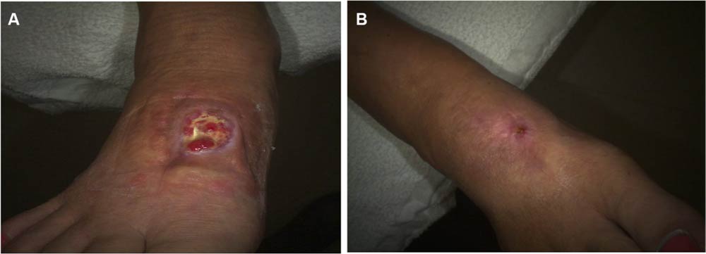
Figure 1 Case 1 surgical wound.
Note: (A) Day 0, 3.3 cm2 ; (B) day 71, 0 cm2.

Case 2 
A 57-year-old patient presented with a left lower extremity VLU on the medial malleolus. The wound had been present for 8 weeks. The treatment thus far consisted of compression and topical antimicrobials. His past medical history included chronic venous insuffciency, peripheral vascular disease, deep vein thrombosis, and hyperlipidemia. On day 0, the wound area measured 0.3 cm . HSAM (2.5 cm x 2.5 cm) was applied and held in place with Steri-Strips™ ( McKesson Medical-Surgical) and compression wrap. On day 7, the wound was completed closed with no drainage (Figure 2).

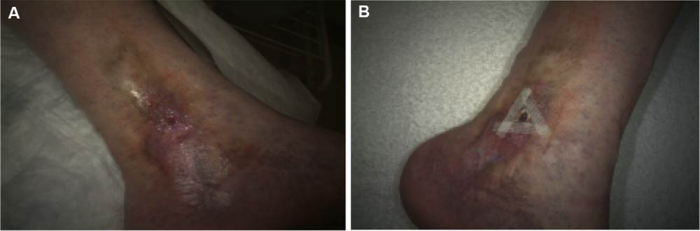
Figure 2 Case 2 venous leg ulcer.
Note: (A) Day 0, 0.3 cm2; (B) day 7, 0 cm2.

Case 3
A 70-year-old patient presented with a VLU on the medial right lower extremity. The wound had been present for 2 weeks. The medical history obtained from this patient revealed hypertension, lymphedema, type II diabetes, and chronic venous insuffciency. At the time of HSAM (2.5 cm × 2.5 cm) application, the wound area measured 1.0 cm . By day 7, the wound area was reduced to 0.4 cm , and by day 14, it achieved complete closure (Figure 3).

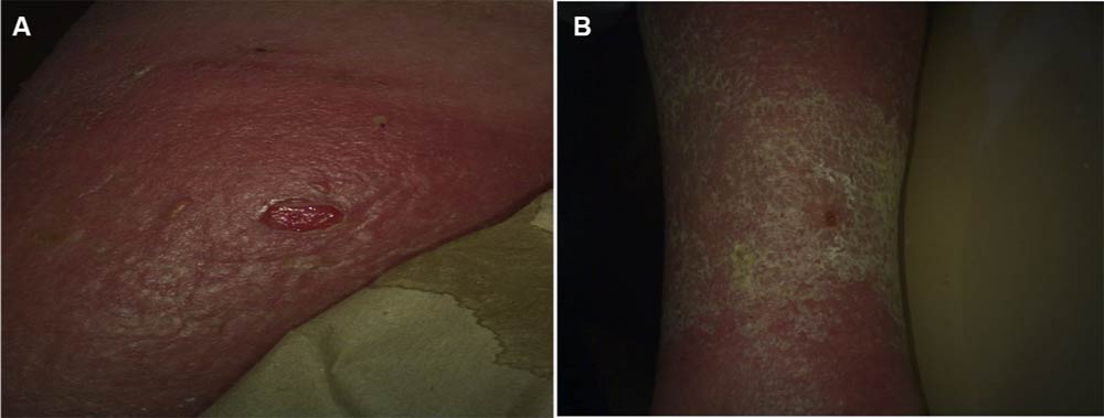
Figure 3 Case 3 venous leg ulcer.
Note: (A) Day 0, 1.0 cm2; (B) day 14, 0 cm2

Discussion
Prompt treatment of chronic ulcers is essential in preventing complications, reducing the cost of care, and lessening the economic burden on the health care system. In clinical studies, amniotic membranes have been shown to be effective in promoting healing in chronic wounds. Prior products consisted of dehydrated products. We postulated that fresh amniotic membrane would demonstrate even greater effect. Prior to embarking on large expensive clinical trials, we tried the fresh amniotic product on a few selected patients. Results from this study suggested that HSAM may be an effective treatment option for management of VLUs and other non-healing wounds. HSAM offers a new treatment alternative to promote healing in chronic wounds. This membrane contains numerous growth factors and cytokines. These growth factors are found to be released over an extended period and result in increased cellular migration, proliferation, and remodeling. Moreover, HSAM may reduce the long-term costs associated with the care of chronic ulcers by increasing the healing rate and lowering the risk of infection and complications This pilot case series was subsequently used to inform larger DFU and VLU trials that are ongoing at the time of this writing.


Original Article – Dovepress


Caring for the Diabetic Foot in Long-Term Care Facilities


By Susan M. Cleveland, BSN, RN, WCC, CDP, NADONA Board Secretary


As a Director of Nursing (DON) in a long-term care facility, do you know where the awareness level of diabetes and its complications is for your staff? Do they realize diabetes doesn’t stop? It is 24/7, 365 days a year. Knowing this reality of diabetes and understanding the disease process may assist with preventing serious health problems such as heart disease, stroke, blindness, kidney disease, and nerve damage that can lead to amputation.


Education is key. We cannot talk about this enough. Assuming someone knows or that common sense will tell people how to take care of these residents is a huge mistake. I don’t believe in common sense; I believe in common knowledge, but it is only common once it is taught. So, teach about diabetes and the care of residents with diabetes often.


Knowing the Risk of Diabetic Foot Ulcers and Diabetic Peripheral Neuropathy
In 2015, 30.3 million Americans, or 9.4% of the population, had diabetes. Approximately 1.25 million American children and adults have type 1 diabetes. The percentage of Americans age 65 and older remains high, at 25.2%, or 12.0 million seniors (diagnosed and undiagnosed).1Diabetic foot ulcer (DFU) is the number one risk factor contributing to non-traumatic foot amputations in persons with diabetes. Limb amputations are preceded by DFUs 58% of the time. The primary risk factor for the DFU is loss of protective sensations or diabetic peripheral neuropathy (DPN) … read more

Toward Eliminating Diabetic Foot Amputation In The Next Generation

David G. Armstrong, professor of surgery at the Keck School of Medicine of USC

Humanity crossed a line in 2009: For the first time in history, more people in the last decade died from non-communicable disease than from all the plagues in the world combined. We’ve entered what science pundit Steve Jones has, in a rather macabre but perceptive manner, dubbed the Age of Decay.


First was the Age of Disaster — 95 percent of human history — when people died from starvation, accidents, violence and cold temperatures. Around 12,000 years ago, we learned to “circle the wagons,” if you will, as a species. As we became agrarian, we moved closer together to store food. A natural byproduct of this was the Age of Disease, when plagues decimated populations. We’re now in the Age of Decay, when the big killers are non-communicable diseases — cancer, cardiovascular disease and diabetes. Pulmonary disease is fourth.


The longer that I have labored in my line of work, the more I realize it is the height of hubris as a clinician to think that I can “fix” anyone. Rather, I think the best thing I can do is help folks move through the world a little bit better. Our goal in this age as clinicians, scientists, policymakers, is to think not about fixing everything but rather to delay decay.


We all rightly take cancer and heart attacks seriously. Diabetes, however, has not risen to that level. If one were an evil deity and wanted to sock it to humanity, one wouldn’t pick something like cancer or a heart attack. Those are often far too dramatic. One would rather choose diabetes: It is silent, sinister and it happens in the background. No one sees it coming. How, then, does diabetes cause amputations?

read more

Diagnosing A Pruritic Skin Lesion In

    A Patient With Diabetes And Edema


A 56-year-old man presented to our institution with the chief complaint of a scaly, pruritic lesion on the lateral aspect of his left lower leg. He would notice some mild bleeding when scratching the area. He denied having any other lesions on his body and there was no previous treatment.


The patient’s past medical history included type 2 diabetes with Charcot arthropathy and neuropathy, hypertension, atrial fibrillation, and deep venous thrombosis. His past surgical history was remarkable for cardiac ablation. The patient is married and does not use tobacco products. He admitted to rare consumption of alcohol.


The patient’s physical exam revealed a man who appeared healthy. The pedal pulses were palpable with capillary refill in less than two seconds to the digits. Mild edema and varicosities were present at the ankle region bilaterally. The neurologic exam with the 10-gram Semmes-Weinstein monofilament showed decreased sensation to the dorsal and plantar aspects of both feet … read more

Ultrasonic debridement system in patients with diabetic foot ulcers

     A case series

Objective:
This study evaluated the use of ultrasonic debridement in patients with diabetic foot ulcers (DFU).


Method:
In this prospective, single-arm, open-label study, all patients with DFUs underwent wound debridement by ultrasonic debridement system (SonicOne OR Ultrasonic debridement system). Wherever possible, the edges were approximated by means of stitches. In other cases, the surgical breach healed by secondary intention, or a partial thickness skin graft (with or without Integra Dermal Regeneration Template or Integra Flowable Wound Matrix) was applied, and subsequently healed by primary intention … read more

Silver dressings improve diabetic wound healing without reducing bioburden

Abstract:
Introduction. Silver dressings are widely used in the treatment of chronic wounds to reduce bacterial bioburden. However, little is known about the mechanism of silver ions on the healing process. In this study, a mouse model of wound healing was used to examine the effect of silver dressings in normal and diabetic wounds.


METHODS:
Two 5-mm full-thickness wounds were created on the dorsal skin of diabetic BKS.Cg- m+/+Leprdb/J mice (experimental group) and wild type C57BL/6 mice (control group), and treated with either a silver or gauze dressing. Measurement of wound areas by digital planimetry demonstrated faster healing in the silver-treated wounds of both diabetic and control mice.


RESULTS:
Quantitative bacterial cultures showed a reduction of bioburden in silver-treated wounds in wild type mice. Unexpectedly, there was no decrease in bioburden in the silver-treated diabetic wounds compared to the control diabetic wounds, despite improved healing in the silver-treated diabetic wounds. Staphylococcus xylosus, a known biofilm producer, was the only bacteria identified in all the wounds. In vitro studies showed S. xylosus produced biofilms faster in higher glucose environments; this may explain the increased bioburden in the wounds in diabetic mice compared to wild type mice.


CONCLUSION:
The results demonstrate improved healing and reduced bioburden in normal wounds with silver dressings. In contrast, silver dressings improved healing in diabetic wounds despite no effect on bioburden, suggesting silver may have beneficial effects in addition to its antimicrobial properties.

Original article from The National Library of Medicine (NLM) 

The Neuropathic Diabetic Foot Ulcer Microbiome Is Associated With Clinical Factors


Nonhealing diabetic foot ulcers (DFUs) are a common and costly complication of diabetes. Microbial burden, or “bioburden,” is believed to underlie delayed healing, although little is known of those clinical factors that may influence microbial load, diversity, and/or pathogenicity. We profiled the microbiomes of neuropathic nonischemic DFUs without clinical evidence of infection in 52 individuals using high-throughput sequencing of the bacterial 16S ribosomal RNA gene. Comparatively, wound cultures, the standard diagnostic in the clinic, vastly underrepresent microbial load, microbial diversity, and the presence of potential pathogens. DFU microbiomes were heterogeneous, even in our tightly restricted study population, but partitioned into three clusters distinguished primarily by dominant bacteria and diversity. Ulcer depth was associated with ulcer cluster, positively correlated with abundance of anaerobic bacteria, and negatively correlated with abundance of Staphylococcus. Ulcer duration was positively correlated with bacterial diversity, species richness, and relative abundance of Proteobacteria, but was negatively correlated with relative abundance of Staphylococcus. Finally, poor glycemic control was associated with ulcer cluster, with poorest median glycemic control concentrating to Staphylococcus-rich and Streptococcus-rich ulcer clusters. Analyses of microbial community membership and structure may provide the most useful metrics in prospective studies to delineate problematic bioburden from benign colonization that can then be used to drive clinical treatment … read more

Corstrata Foot Ulcer Prevention Solution

  Named Among Semifinalists For T1D Exchange 2018 Diabetes Innovation Challenge


Corstrata provides new tech empowered diabetic foot ulcer prevention to improve patient lives.


CORSTRATA, a provider of digital healthcare IT solutions and services for wound management, announced today that the company’s Diabetic Foot Ulcer & Amputation Prevention solution was named as a semifinalist in the T1D Exchange 2018 Diabetes Innovation Challenge. Corstrata was one of 30 semifinalists chosen from academic researchers and early stage companies from around the world that submitted entries for solutions to support advanced diabetes care. T1D Exchange is a nonprofit research and collaboration organization dedicated to accelerating novel treatments to improve the care of people living with type 1 diabetes (T1D).


Diabetic foot ulcers (DFUs) are a common, limb-threatening, and expensive complication of diabetes. Of the 29M people with diabetes in the U.S., 1.7M suffer with one or more DFUs annually and 80K of these diabetics ultimately require an amputation. The risk of death at 5 years for DFU patients is 2.5 times as high as the risk for a patient with diabetes without a foot ulcer.


Corstrata offers a technology-enabled care management solution for diabetics at risk for formation of costly diabetic foot ulcers and related amputations. Using a “smart” mat to detect potential ulcers, Corstrata’s wound specialists engage with the patient through a mobile engagement app for timely intervention and prevention of ulcers. Corstrata’s Diabetic Foot Ulcer & Amputation Prevention Program serves as an outsourced end-to-end technology-enabled solution for value-based organizations and payers … read more

Biofilm infections between Scylla and Charybdis

Interplay of host antimicrobial peptides and antibiotics


Purpose: The aim of this study is to improve the anti-biofilm activity of antibiotics. We hypothesized that the antimicrobial peptide (AMP) complex of the host’s immune system can be used for this purpose and examined the assumption on model biofilms.


Methods: FLIP7, the AMP complex of the blowfly Calliphora vicina containing a combination of defensins, cecropins, diptericins and proline-rich peptides was isolated from the hemolymph of bacteria-challenged maggots. The complex interaction with antibiotics of various classes was studied in biofilm and planktonic cultures of Staphylococcus aureusEscherichia coliPseudomonas aeruginosaKlebsiella pneumoniae and Acinetobacter baumannii by the checkerboard method using trimethyl tetrazolium chloride cell viability and crystal violet biofilm eradication assays supplemented with microscopic analysis.


Results: We found that FLIP7 demonstrated: high synergy (fractional inhibitory concentration index <0.25) with meropenem, amikacin, kanamycin, ampicillin, vancomycin and cefotaxime; synergy with clindamycin, erythromycin and chloramphenicol; additive interaction with oxacillin, tetracycline, ciprofloxacin and gentamicin; and no interaction with polymyxin B. The interaction in planktonic cell models was significantly weaker than in biofilms of the same strains. The analysis of the dose–effect curves pointed to persister cells as a likely target of FLIP7 synergistic effect. The biofilm eradication assay showed that the effect also caused total destruction of S. aureus and E. coli biofilm materials. The effect allowed reducing the effective anti-biofilm concentration of the antibiotic to a level well below the one clinically achievable (2–3 orders of magnitude in the case of meropenem, ampicillin, cefotaxime and oxacillin).

read more

Complications in Chronic Wound Healing and

Associated Interventions


Overview of Chronic Wounds
Chronic non-healing wounds affect millions of patients each year and contribute significantly to their morbidity and mortality. These wounds have a substantial impact because of their economic burden and the significant effect on the reduction in quality of life, as well as the increased risk of death for those patients affected by them. A 2014 study of Medicare data showed that chronic non-healing wounds and associated complications affect nearly 15% or 8.2 million Medicare beneficiaries. The study also estimated the cost to treat these wounds at between $28.1 billion and $31.7 billion annually. The highest costs were associated with infected or reopened surgical wounds, and outpatient care had the highest site-of-service costs. In addition to being older, most of these patients have obesity and diabetes. Underlying causes often include diabetic foot ulcers, venous leg ulcers, arterial insufficiency, and pressure ulcers. The list of complications contributing not only to chronicity but also to further deterioration is quite lengthy.


Specific Wound Healing Complications and Interventions
The prevalence of chronic wounds and their complications has not been well documented in the literature. However underappreciated they may be, the complications associated with chronic wounds increase the cost—both financial and personal—to the individuals with these wounds.Although not an all-inclusive list, some of the more common complications include infection, tissue necrosis and gangrene, periwound dermatitis, periwound edema, osteomyelitis, hematomas, and dehiscence. Our purpose is to discuss the presentation of each and potential interventions … read more

Initiative Launches Web Portal for Medical Students

Free resources provide education on chronic wound management


In an effort to further prepare medical professionals about the proper management and treatment of patients with wounds, HMP, a leading healthcare event and education company, today announced the launch of a new web portal designed exclusively for medical students as part of its Why Wound Care? (WWC) initiative.


Created in 2015, the Why Wound Care? initiative informs medical and nursing students, recent graduates, and faculty about rewarding careers in wound care while offering educational resources to supplement current academic curricula where wound care education may be limited.


With the development of the new portal, medical students and faculty now have access to the following complimentary, evidence-based wound care resources:


-Sixteen video modules covering the fundamentals of wound care, including
Burns, Surgical Wound Closure, Wound Epidemiology, Pressure Injuries, Diabetic Foot Ulcers … read more


Roles of alternative activation of macrophages phenotypes in normal wound healing

Normal wound healing process is characterized by highly organized controlled overlapping phases including haemostasis, inflammation, proliferation, and remodeling; through which acute wounds come to a complete healing with predictable time frame (Schreml et al, 2010). These phases are orchestrated by the interaction of different cell types and biochemical components to regulate and accomplish the different wound healing process such as coagulation, chemotaxis, phagocytosis, synthesis of extracellular matrix components, angiogenesis, epithelial migration, and remodeling components (Velnar et al, 2009). Components of the main cellular wound healing process include macrophages, keratinocytes, endothelial cells, fibroblast, neutrophils, and lymphocytes. These components are recruited, stimulated and activated according to their role in the healing process by which specific generic cytokines, growth factors, chemokines, and respective receptors are created to achieve physiological wound healing of skin wounds (Schreml et al, 2010) … read more

Meeting Report: ‘Raising the Bar’- creating a better tomorrow

This article is based on the proceedings of the one-day ‘Raising the Bar’ conference in Singapore held by Wounds International and supported by an educational grant from Urgo International on October 15, 2017. The day provided more than 100 clinicians from around Asia with an exceptional opportunity to gain insights and best practice recommendations from a faculty of local and regional wound care specialists, supported by visions from global practitioners … read more

The Effect of Foot Exercises on Wound Healing

in Type 2 Diabetic Patients With a Foot Ulcer: A Randomized Control Study


PURPOSE: The purpose of this study was to investigate the effect of foot exercises on wound healing in type 2 diabetic patients with a diabetic foot ulcer.


DESIGN: Prospective, randomized controlled study.


SUBJECT AND SETTINGS: Sixty-five patients from an outpatient clinic with grade 1 or 2 ulcers (Wagner classification) who met study criteria agreed to participate; 60 patients completed the study and were included in the final analysis. Subjects were followed up between February 2014 and June 2015.


METHODS: Subjects were recruited by the researchers in the clinics where they received treatment. Subjects were randomly allocated to either the control or intervention group. Data were collected using investigator-developed forms: patient information form and the diabetic foot exercises log …. read more

Bacteria can pass on memory to descendants, researchers discover

Led by scientists at UCLA, an international team of researchers has discovered that bacteria have a “memory” that passes sensory knowledge from one generation of cells to the next, all without a central nervous system or any neurons.


“This is a huge surprise to us and to the field,” said Gerard Wong, a professor of bioengineering and of chemistry and biochemistry, member of the California NanoSystems Institute at UCLA and one of the study’s senior authors.


These findings are a major step toward understanding hard-to-treat infections caused by bacterial biofilms in people with cystic fibrosis.


The team studied a strain of bacteria called Pseudomonas aeruginosa that forms biofilms in the airways of people with cystic fibrosis and causes persistent infections that can be lethal. Bacterial biofilms can also form on surgical implants, like an artificial hip; when they do, they can cause the implant to fail. Bacterial biofilms are composed of genetically identical bacteria cells that can colonize nearly any surface and form communities in which single cells organize and cooperate … read more


Watch How Engineering Students Step Up with Imager for Diabetics

Patients with diabetes must guard against nerve damage disorders that can numb feeling in their extremities. Engineering students developed a device to help patients with diabetes easily monitor their feet for cuts or other injuries.

Engineering students developed a device to help patients with diabetes easily monitor their feet for cuts or other injuries.


A simple device developed by Rice University students can help this population detect early signs of foot ulceration that, left untreated, could endanger their health and lead to amputation.The inspection device is likened to a foot stand one might see in a shoe store but with the addition of a clear plastic top, cameras, lights, a mirror and electronics that allow people to easily examine their feet.


The initial design attached a camera on a gooseneck to the end of a telescoping stick that patients could manipulate to manually inspect their feet … read more


Digital wound care provider nabs $11.6M in funding

A Toronto startup, looking to revolutionize wound care, just got a big boost in its expansion efforts.

Swift Medical recently announced that it has earned $11.6 million in funding, which will help to fuel its goal of being used across North America. The smartphone-based care aid helps clinicians to measure wounds, without touching the resident or using extra accessories. Already, it’s been adopted in more than 1,000 long-term care and other types of facilities.

“This growth funding enables us to expand our reach and bring Swift’s solution to every bedside in every hospital and care facility,” Carlo Perez, co-founder and CEO of Swift Medical, said in a  press release. “There are more patients worldwide suffering from chronic wounds than from lung cancer, breast cancer, colon cancer and leukemia combined. By augmenting the abilities of clinicians and facility administrators to deliver the best possible wound care management, we’re helping them heal over 10,000 patients a month. And we’re just getting started.”

read more

The use of Prontosan® in combination with Askina® Calgitrol®

An independent case series

Many patients with chronic wounds will develop infection (Landis et al, 2007; Sibbald et al, 2011). Worldwide consensus on the specific use of silver antimicrobials recommends that silver dressings should be used initially for a ‘two-week challenge’ (Wounds International, 2012). Sixteen different individual case studies were carried out to evaluate the efficacy of a biofilm remover/cleanser in gel form, Prontosan® (B Braun), together with the use of an ionic releasing silver alginate, Askina® Calgitrol® Paste (B Braun) or Askina® Calgitrol® Thin (B Braun), when used on infected wounds. This study was completed in an advanced wound management centre in Pretoria, South Africa, during 2016.  Selection criteria included wounds showing clinical signs of infection with delayed healing for more than 2 weeks. The study results showed that 50% of the wounds’ clinical signs of infection resolved within the 2-week antimicrobial challenge and by week 3, 81% of all clinical signs resolved. Ninety-three per cent of the wounds had improved wound progress and healing .. read more


Reduction of 50% in Diabetic Foot Ulcers With Stem Cells

MUNICH — Local injection of mesenchymal stem cells derived from autologous bone marrow shows promise in healing recalcitrant neuropathic diabetic foot ulcers, a novel study from Egypt shows.


Presenting the results at the European Association for the Study of Diabetes (EASD) 2016 Annual Meeting, Ahmed Albehairy, MD, from Mansoura University, Egypt, said: “In patients who received the mesenchymal stem cells, ulcer reduction was found to be significantly higher compared with patients on conventional treatment after both 6 weeks and 12 weeks of follow-up. This is despite the fact that initial ulcer size was larger in the stem-cell–treated group.”


After 6 weeks, median ulcer reductions were 49.9% and 7.67% (P = .001) in stem-cell–treated and control groups, respectively, and after 12 weeks, median ulcer reductions were 68.24% and 5.27% (P = .0001). Complete healing was achieved in one case in the mesenchymal stem cell–treated group.


“The healing mechanism may be due to the pure effect of injected mesenchymal stem cells, which is due to the ability of these cells to simulate angiogenesis in the wound bed, decrease excessive inflammation, and suppress scarring,” explained Dr Albehairy … read more

Clinical Challenges in Diagnosing Infected Wounds

Given the impact of infection on delayed wound healing, determining the presence of colonization and infection is imperative to achieving healed outcomes. Chronic wounds are always contaminated, and timely implementation of management and treatment interventions is a key component of the plan of care.


Diagnosis of infection can be a very challenging task to say the least, and it is further complicated by the presence of biofilms for which no diagnostic tool is currently available. If not addressed in a timely manner, these local infections can become systemic, leading to sepsis, multiple organ failure, and death. The first steps are a complete and thorough history and a physical examination of the whole patient, not just the patient’s wound, while taking into account both primary and secondary findings to understand the host response.


Having a thorough understanding of the principles of chronic wound care and of the current diagnostic modalities available is essential to the improvement of clinical outcomes and cost reduction related to the complication of wound infection. Our focus is on the challenges to diagnosing wound infection, including accurately determining risk factors, differentiating colonization from infection, and understanding the gold standard for diagnosing wound infection … read more

ARANZ Medical Launches Two New Silhouette Image Capture Products


CHRISTCHURCH, New ZealandMarch 28, 2018 /PRNewswire/ — Two new smart-phone based imaging products, SilhouetteLite+ and SilhouetteLite, recently launched by ARANZ Medical Limited have been adopted by Promore Pharma AB (Solna, Sweden) for a multi-site clinical trial, and by New Zealand’s Capital and Coast District Health Board for their community-based nurses.

Easy-to-use, SilhouetteLite+ and SilhouetteLite apps are highly portable, yet deliver robust accuracy and image consistency. The measurement and documentation data collected is securely synchronized in ARANZ Medical’s proprietary, cloud database, SilhouetteCentral, for ease of analysis and reporting.


Promore Pharma purchased 20 SilhouetteLite+ units for their two-year venous leg ulcer study. Chief Scientific Officer, Dr. Margit Mahlapuu, stated, “The SilhouetteLite+ app and documentation system is easy for us to use across multiple locations. With it, we can ensure that the data we collect at each site is not only consistent and secure, but also easily accessed for analysis and reporting.”

read more


Wound Care in Crisis

Wound Care’s Newest Buzzword and Slogan


Every so often, in my readings of newspapers, articles, and while pleasure reading, or during conversations with friends and colleagues, I come across a word that smacks me right in the kisser. I was listening to a news commentary and this new buzzword nonchalantly emanated from the speaker’s lips. It took me but a second before I realized what an insidiously powerful little devil it was. The speaker droned on about the current political scenario, the state of medical care, and then, in Judge Wapner-like reckoning described his presumed foes points as “counterintuitive.” This unusual word “counterintuitive” has a definition that is both painfully simple and thought-provokingly powerful. Intuition is simply the perception of something using common sense. Adding “counter” to it moves it into the realm of mystery and suspense. Counter intuitive, something that is unlikely to be found correct when assessed and evaluated.


The problem is that the meaning of counterintuitive is usually in the eyes of the beholder or the “be-dabbler” in the case of wound care. There are far too many things done in an incorrect, non-evidence-based, un-best-practice-based manner in the profession that I love so dearly. The problem is that identifying something as counterintuitive means that you have to have some semblance of intuition regarding the issue or in other words, half an idea of what you are talking about. The real issue, however, is that the care you provide needs to be self-scrutinized, self-evaluated, and dare I say it, reviewed by your peers and those you work intimately with who will provide you an honest, unbiased opinion regardless of whether you like it or not.


I received a call from a patient in a long-term care facility to resume care for him. Once at home and doing well, he had fallen into the black hole of the local medical center and then was unceremoniously dumped into a long term care facility. The astute wound care NP running their wound “lack of care” program wrote a War and Peace worthy history and physical and then documented his venous insufficiency, venous insufficiency ulcers, mild secondary lymphedema and his obesity with debility … read more


Bioelectrical Impedance Assessment of Wound Healing

Objective assessment of wound healing is fundamental to evaluate therapeutic and nutritional interventions and to identify complications. Despite availability of many techniques to monitor wounds, there is a need for a safe, practical, accurate, and effective method. A new method is localized bioelectrical impedance analysis (BIA) that noninvasively provides information describing cellular changes that occur during healing and signal complications to wound healing. This article describes the theory and application of localized BIA and provides examples of its use among patients with lower leg wounds. This promising method may afford clinicians a novel technique for routine monitoring of interventions and surveillance of wounds.


Wound healing is a dynamic, interactive cascade of molecular, cellular and biochemical processes. Despite accumulating knowledge of the biology of wound healing, the estimated annual cost of treating wounds exceeds $20 billion in the United States, particularly for wounds of the lower body. Although many therapeutic interventions are utilized to treat wounds, physician decisions are hampered by the lack of objective and convenient methods to monitor treatment effects and to assess wound healing. Contemporary methods have limitations including cost, time commitment, reliability, and accuracy. Thus, the need persists for an objective, suitable, and practical method to assess wound healing.


Assessment of the effectiveness of treatment to foster wound healing is a complex and broad field. Traditional methods estimate the dimensions of wounds including surface area and volume. Physiological approaches utilize molecular and biochemical indicators that provide less subjective information. Because successful wound healing is a dynamic process that integrates physiological and biochemical factors and mechanisms, reliance on a single aspect of the process may be inadequate. This article describes the use of localized bioelectrical impedance analysis (BIA) measurements to monitor cellular processes involved in wound healing …  read more

Dr. Tim Lu – Biofilms and Phage Therapy

This 11 minute film is excerpted from an interview with Dr. Tim Lu, who is an expert in characterizing & eliminating biofilms with phage therapy. He offers some insightful ways to describe complex biofilms and their connection to antibiotic resistance.


Interview excerpts & videos with Bacterial Biofilm Experts (doctors & researchers):
www.biofilmcommunity.org

Smart socks aim to catch diabetic foot problems early

SAN FRANCISCO (KGO) — For the millions of Americans living with diabetes, a new tech product offers early warning against the threat of losing a foot or a leg to the disease.


“I have very little sensation on the bottom of my feet,” explained Marc Fairman, who’s type 1 diabetic. Fairman will never take his feet for granted again. In 2012, he developed a foot ulcer that got out of control.


“I sought several opinions — surgeons and podiatrists — and several told me i would probably lose my foot,” he said. For 30 million Americans with diabetes, UCSF surgery professor Michael Conte said it’s a very real danger.


read more

Berlin doctor lauded for leg amputation research

BERLIN – A local doctor has received international honors for a scientific paper about a new scoring system he helped create to evaluate the risks of leg amputations.


Dr. Eric Newgent, medical director of sleep medicine at ThedaCare Medical Center-Berlin, wrote the article for The Journal of Wound Care with Dr. Michael Miller, a general surgeon and full-time wound care specialist in Indiana, according to ThedaCare … read more


A comprehensive scoring system to evaluate patient-centered risk factors regarding lower extremity amputation


Assessment and management of foot ulcers

for people with diabetes.


Major Recommendations

The levels of evidence supporting the recommendations (Ia, Ib, IIa, IIb, III, IV) are defined at the end of the “Major Recommendations” field.


Practice Recommendations


Assessment


Recommendation 1.0


Obtain a comprehensive health history and perform physical examination of affected limb(s).

(Level of Evidence = Ib–IV)


Recommendation 1.1


Identify the location and classification of foot ulcer(s) and measure length, width, and depth of woundbed.

read more